Perthes Disease X Ray - Perthes Disease X Ray Page 3 Line 17qq Com - This video describes the pathogenesis (natural history of disease) and x ray features of legg calve perthes disease (avascular necrosis of hip joint in.
Perthes Disease X Ray - Perthes Disease X Ray Page 3 Line 17qq Com - This video describes the pathogenesis (natural history of disease) and x ray features of legg calve perthes disease (avascular necrosis of hip joint in.. Technetium bone scan or mri scanning can. (2017) clinical cases in mineral and bone metabolism. During healing, the socket part of the joint can serve as a mold to help the fragmented femoral head retain its round shape. Of function, clinical parameters and radiological changes. Normal x rays in perthes disease.
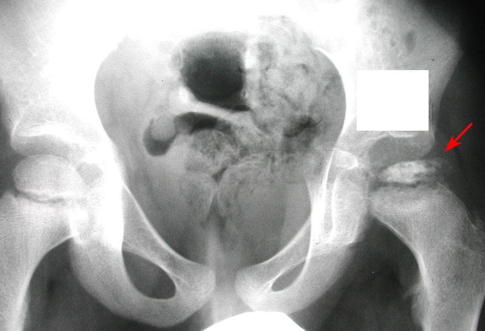
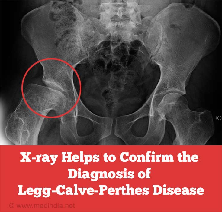
The aim of surgical intervention is to cover the femoral head as completely. A more detailed differentiation of waldenström's classification, especially in early and late fragmentation stages. Note the slight widening of the left hip joint meadows c, monsell f, ramanan av. For this molding to work, the femoral head must sit snugly within. Due to the lack of blood flow, the bone dies (osteonecrosis or avascular necrosis) and stops growing.
Perthes Disease Legg Calve Perthes Orthoinfo Aaos from orthoinfo.aaos.org Note the slight widening of the left hip joint meadows c, monsell f, ramanan av. Perthes disease is a condition that affects the hip in children between the ages of four and eight. Nord guide to rare disorders. Your doctor may request a bone scan, ultrasound or mri scan. Perthes' disease usually affects children between the ages of three and eleven years. A bone scan or mri may be useful if the diagnosis is. Perthes disease is relatively uncommon and in western populations has an incidence approaching 5 to 15:100,000. A study of 610 children under 12 years of age at disease onset.
It is due to avascular necrosis of the femoral head, specifically the femoral epiphysis.
(2017) clinical cases in mineral and bone metabolism. A more detailed differentiation of waldenström's classification, especially in early and late fragmentation stages. A bone scan or mri may be useful if the diagnosis is. Legg calvé perthes disease is a condition characterized by a temporary loss of blood supply to the top of the femur. For this molding to work, the femoral head must sit snugly within. During healing, the socket part of the joint can serve as a mold to help the fragmented femoral head retain its round shape. The patient was seen again at the age of 8 years. Perthes disease is a rare kind of disability, primarily affecting the hip joint of children. A study of 610 children under 12 years of age at disease onset. Note that the femoral head is completely contained inside the hip cup. Perthes disease (lcpd) are based on subjective measures. Note the slight widening of the left hip joint meadows c, monsell f, ramanan av. Impaired blood supply to the femoral head causes bone infarction.
Due to the lack of blood flow, the bone dies (osteonecrosis or avascular necrosis) and stops growing. A bone scan or mri may be useful if the diagnosis is. During healing, the socket part of the joint can serve as a mold to help the fragmented femoral head retain its round shape. Nord guide to rare disorders. Perthes disease is a condition that affects the hip in children between the ages of four and eight.
Legg Calve Perthes Disease Childhood Bone Disorder from images.medindia.net Coronavirus disease programme online course: A study of 610 children under 12 years of age at disease onset. Due to the lack of blood flow, the bone dies (osteonecrosis or avascular necrosis) and stops growing. Physical measurements of the thigh muscles and exams to assess the kids' range of motion are also performed as part of diagnosis. It is more common in boys than in girls. Perthes disease is a rare kind of disability, primarily affecting the hip joint of children. Nord guide to rare disorders. A bone scan or mri may be useful if the diagnosis is.
A more detailed differentiation of waldenström's classification, especially in early and late fragmentation stages.
Physical measurements of the thigh muscles and exams to assess the kids' range of motion are also performed as part of diagnosis. Perthes disease (lcpd) are based on subjective measures. A bone scan or mri may be useful if the diagnosis is. This may mean careful explanation of the procedure and even immobilizing a child to decrease movement. Your doctor may request a bone scan, ultrasound or mri scan. Normal x rays in perthes disease. (2017) clinical cases in mineral and bone metabolism. Of function, clinical parameters and radiological changes. Due to the lack of blood flow, the bone dies (osteonecrosis or avascular necrosis) and stops growing. A more detailed differentiation of waldenström's classification, especially in early and late fragmentation stages. The aim of surgical intervention is to cover the femoral head as completely. Related online courses on physioplus. It is due to avascular necrosis of the femoral head, specifically the femoral epiphysis.
Note that the femoral head is completely contained inside the hip cup. Normal x rays in perthes disease. A study of 610 children under 12 years of age at disease onset. This may mean careful explanation of the procedure and even immobilizing a child to decrease movement. (2017) clinical cases in mineral and bone metabolism.
Legg Calve Perthes Disease Wikipedia Republished Wiki 2 from upload.wikimedia.org This may mean careful explanation of the procedure and even immobilizing a child to decrease movement. Natural evolution of perthes disease: It is due to avascular necrosis of the femoral head, specifically the femoral epiphysis. (2017) clinical cases in mineral and bone metabolism. Coronavirus disease programme online course: Physical measurements of the thigh muscles and exams to assess the kids' range of motion are also performed as part of diagnosis. Impaired blood supply to the femoral head causes bone infarction. Perthes disease is relatively uncommon and in western populations has an incidence approaching 5 to 15:100,000.
Perthes disease (lcpd) are based on subjective measures.
Coronavirus disease programme online course: It is due to avascular necrosis of the femoral head, specifically the femoral epiphysis. It is more common in boys than in girls. A study of 610 children under 12 years of age at disease onset. Nord guide to rare disorders. Normal x rays in perthes disease. (2017) clinical cases in mineral and bone metabolism. This may mean careful explanation of the procedure and even immobilizing a child to decrease movement. Legg calve perthes disease sometimes called perthes disease or coxa plana, or avascular necrosis of the hip, is a bone disorder that affects the hips of children. The aim of surgical intervention is to cover the femoral head as completely. Natural evolution of perthes disease: Perthes disease is relatively uncommon and in western populations has an incidence approaching 5 to 15:100,000. This video describes the pathogenesis (natural history of disease) and x ray features of legg calve perthes disease (avascular necrosis of hip joint in.
Normal x rays in perthes disease perth. Perthes disease is a condition that affects the hip in children between the ages of four and eight.


0 Komentar